- Home
- 图像长廊
Select an application or technique:
Latest Images
超快速原子力显微镜
光镊-原子力显微镜联用以及先进光学
生命科学
聚合物
纳米科学
电、磁、热等
细胞力学与细胞粘附
单分子力谱
纳米操纵与蚀刻
Raman, TERS 以及 SNOM
原子力显微镜全自动力谱仪光镊/光阱细胞粘附力/细胞力学测试组件
NanoWizard® BioScience AFM
-
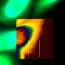
 Living A549 cells - Correlative AFM and STED
 -
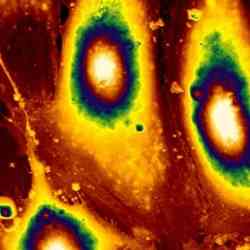
 Living Vero cells
 -
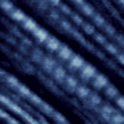
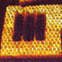 DNA Origami at 150 lines/sec
 -
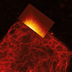
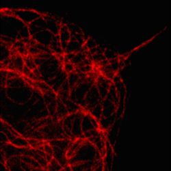
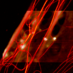
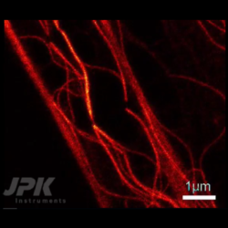 VIDEO - Real-time rapture of microtubules - Correlative AFM and STED measurements
 -
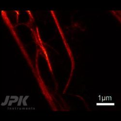 VIDEO - Real-time bending of microtubules - Correlative AFM and STED measurements
 -
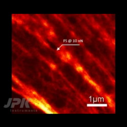 VIDEO - Stimulation of living fibroblast cells - Correlative AFM and STED measurements
 -
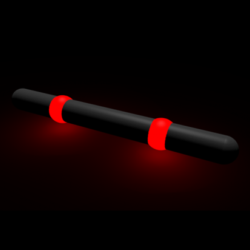 Nanoruler - AFM and STED
 -
 Living A549 cells - Simultaneous AFM and STED
 -
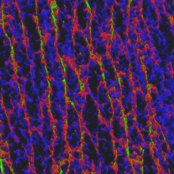
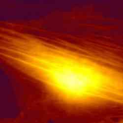
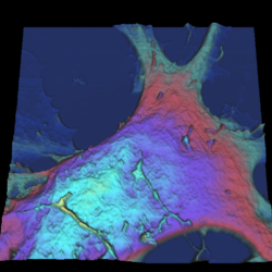 Astrocytes
 -
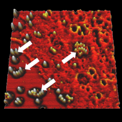
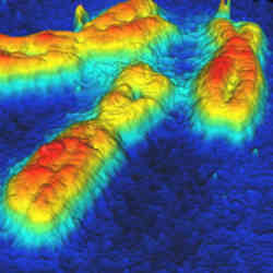
 Imaging of bacteria S-layer with QI™
 -
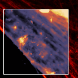 Simultaneous AFM and STED of Living fibroblasts - Actin Filament Imaging
 -
 Simultaneous AFM and STED of Living fibroblasts - Microtubule Imaging
 -
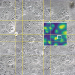
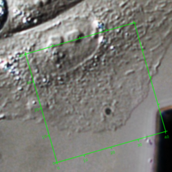
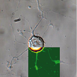
 Cell/particle interaction - AFM with confocal microscopy
 -
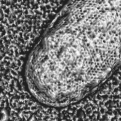
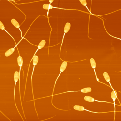 High-resolution imaging on sperm
 -
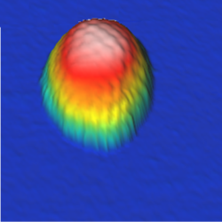
 Living CHO cell
 -
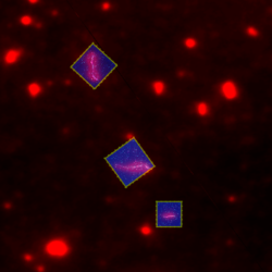 Rad51 proteins bound to DNA - AFM with fluorescence microscopy
 -
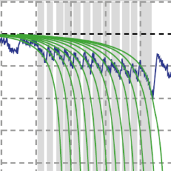 Fibronectin unfolding
 -
 DNA at -25 °C
 -
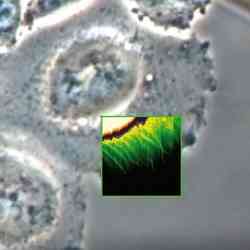 CHO cell - AFM with phase contrast
 -
 QI™ DNA - Major and minor grooves
 -
 Recognition microscopy on biotin bead
 -
 Recognition on living keratinocyte cells
 -
 DNA origami - faceman
 -
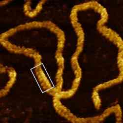
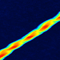
 Twisted insulin fibrils
 -
 Living CHO cells - AFM with fluorescence microscopy
 -
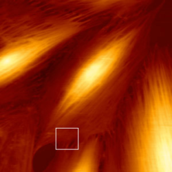
 Tendon tissue
 -
 Herpes Simplex Viruses
 -
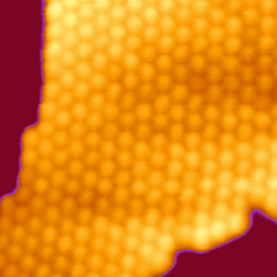
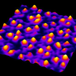 Bacteriorhodopsin membrane - QI™ mode
 -
 Cell division E-coli bacteria
 -
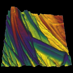
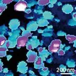
 Human dental enamel
 -
 VIDEO - Live CHO cell dynamics
 -
 Tomato Bushy Stunt Virus
 -
 Plasmid DNA imaged in HyperDrive™ mode in buffer
 -
 KPG7 cell dynamics
 -
 Living Candida albicans - AFM with phase contrast
 -
 HeLa cell in buffer - AFM with STORM
 -
 Melting of lipid domains in buffer
 -
 Collagen in liquid, 70Hz line rate
 -
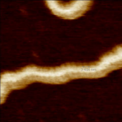
 Lambda phage DNA in liquid
 -
 Bacteriorhodopsin in buffer
 -
 Living fibroblast cell - AFM with phase contrast
 -
 Desulfobulbus bacterial cells
 -
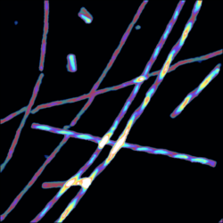
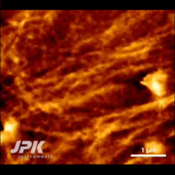
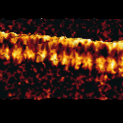
 Collagen fibres
 -
 Pea starch granules
 -
 Herpes Simplex Virus
 -
 Living Cyanobacterium
 -
 Living Escherichia coli bacteria
 -
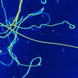
 Living dorsal root ganglion cells - AFM with DIC
 -
 Twisted amyloid fibrils
 -
 Collagen – AFM with phase contrast
 -
 Glucagon fibre
 -
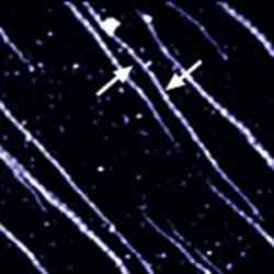
 Fibrillin microfibrils
 -
 Malaria infected red blood cells
 -
 L929 cell filipodia
 -
 Single waste water bacterium
 -
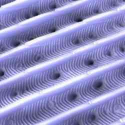
 OmpF protein crystal
 -
 Lipid bilayer - AFM with confocal microscopy
 -
 MDCK cells - AFM with confocal microscopy
 -
 Moth's eye
 -
 Human lymphocyte chromosomes
 -
 Unfixed collagen in buffer
 -
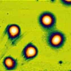
 Ptk2 cells - AFM with fluorescence microscopy
 -
 Living fibroblast cells – AFM with phase contrast and fluorescence
 -
 Living fibroblast cell
 -
 Lipid vesicles
 -
 Moth wing scale
 -
 Collagen manipulation
 -
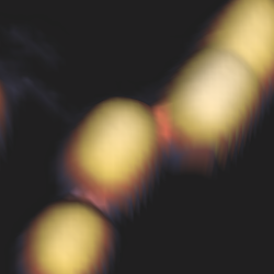
 Gold clusters in water
 -
 REF52 cells – AFM with fluorescence
 -
 High-resoution image of HPI layer