- Home
- 图像长廊
Select an application or technique:
Latest Images
超快速原子力显微镜
光镊-原子力显微镜联用以及先进光学
生命科学
聚合物
纳米科学
电、磁、热等
细胞力学与细胞粘附
单分子力谱
纳米操纵与蚀刻
Raman, TERS 以及 SNOM
原子力显微镜全自动力谱仪光镊/光阱细胞粘附力/细胞力学测试组件
光镊-原子力显微镜联用以及先进光学
Superresolution Microscopy (STED, STORM, PALM)
-
 NanoWizard® BioScience AFM
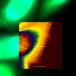
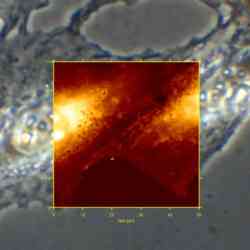
Living A549 cells - Correlative AFM and STED -
 NanoWizard® BioScience AFM
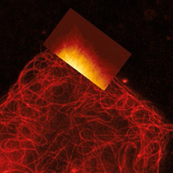
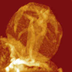
VIDEO - Real-time rapture of microtubules - Correlative AFM and STED measurements -
 NanoWizard® BioScience AFM
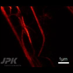
VIDEO - Real-time bending of microtubules - Correlative AFM and STED measurements -
 NanoWizard® BioScience AFM
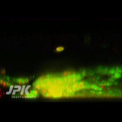
VIDEO - Stimulation of living fibroblast cells - Correlative AFM and STED measurements -
 NanoWizard® BioScience AFM
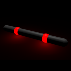
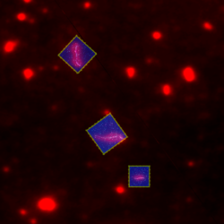
Nanoruler - AFM and STED -
 NanoWizard® BioScience AFM
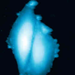
Living A549 cells - Simultaneous AFM and STED -
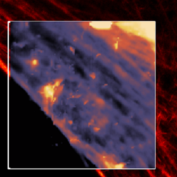 NanoWizard® BioScience AFM
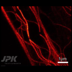
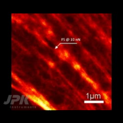
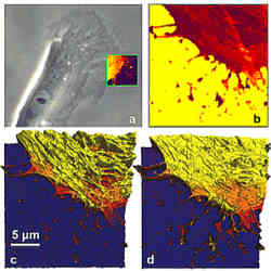
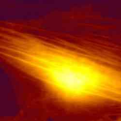
Simultaneous AFM and STED of Living fibroblasts - Actin Filament Imaging -
 NanoWizard® BioScience AFM
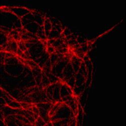
Simultaneous AFM and STED of Living fibroblasts - Microtubule Imaging -
 NanoWizard® BioScience AFM
HeLa cell in buffer - AFM with STORM
Confocal Microscopy, FCS, FLIM, TIRF, IRM
-
 NanoWizard® NanoOptics AFM
Simultaneous AFM and FLIM measurements -
 NanoWizard® BioScience AFM
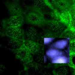
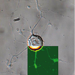
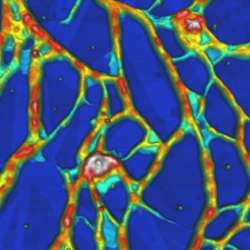
Cell/particle interaction - AFM with confocal microscopy -
 NanoTracker™ 2
Recording of a confocal z-stack -
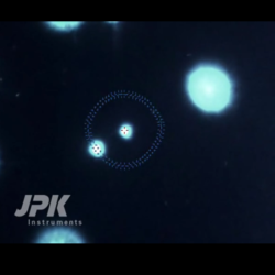
 NanoTracker™ 2
Confocal scanning combined with optical particle manipulation -
 NanoWizard® BioScience AFM
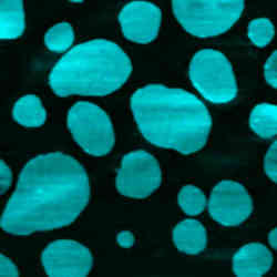
Lipid bilayer - AFM with confocal microscopy -
 NanoWizard® BioScience AFM
MDCK cells - AFM with confocal microscopy -
 NanoWizard® Sense AFM
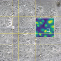
SAOS cells - AFM with confocal microscopy
Fluorescence Microscopy
-
 NanoWizard® BioScience AFM
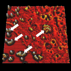
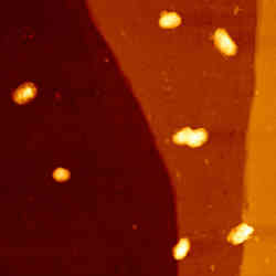
Rad51 proteins bound to DNA - AFM with fluorescence microscopy -
 NanoWizard® BioScience AFM
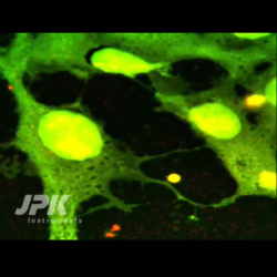
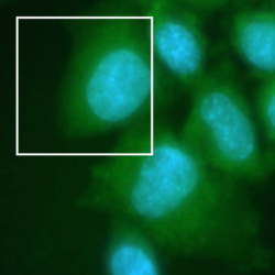
Living CHO cells - AFM with fluorescence microscopy -
 NanoTracker™ 2
Fluorescence filter change during trap manipulation -
 NanoTracker™ 2
Fluorescent microtubule manipulation -
 NanoWizard® NanoScience AFM
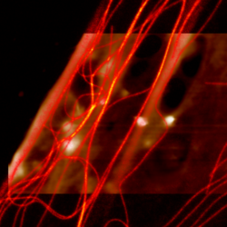
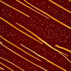
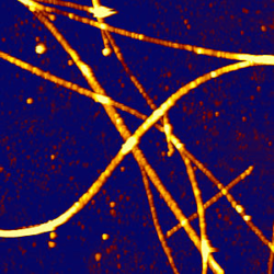
Hexaphenyl nanofibers - AFM with fluorescence microscopy -
 NanoWizard® NanoScience AFM
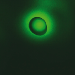
Fluorescent polymer spheres - AFM with fluorescence microscopy -
 NanoWizard® BioScience AFM
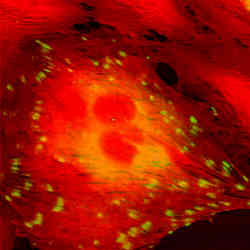
Ptk2 cells - AFM with fluorescence microscopy -
 NanoWizard® Sense AFM
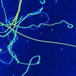
Microtubules – AFM with fluorescence -
 NanoWizard® BioScience AFM
REF52 cells – AFM with fluorescence
Phase Contrast and DIC
-
 NanoWizard® BioScience AFM
Living Vero cells -
 NanoWizard® BioScience AFM
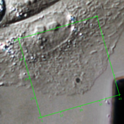
Living CHO cell -
 NanoWizard® BioScience AFM
CHO cell - AFM with phase contrast -
 NanoWizard® BioScience AFM
Living fibroblast cell - AFM with phase contrast -
 NanoWizard® BioScience AFM
Living dorsal root ganglion cells - AFM with DIC -
 NanoWizard® BioScience AFM
Collagen – AFM with phase contrast -
 NanoWizard® NanoScience AFM
Polyelectrolyte shell - AFM with DIC -
 NanoWizard® Sense AFM
SAOS chondrocyte cells – AFM with phase contrast -
 NanoWizard® BioScience AFM
Living fibroblast cells – AFM with phase contrast and fluorescence
Upright Microscopy
-
 BioMAT™ Workstation
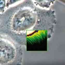
J-aggregates -
 BioMAT™ Workstation
Paracoccus Seriniphilus bacteria -
 BioMAT™ Workstation
Living CHO on gold electrode -
 BioMAT™ Workstation
Cow tooth – etched - AFM with upright microscopy -
 BioMAT™ Workstation
Bacteria on pyrite surface - AFM with upright fluorescence microscopy -
 BioMAT™ Workstation
Mouse cerebellum tissue - AFM with upright fluorescence microscopy